Reading: Haines, Ch. 23, pp 341-346; Ch. 24; Ch.25, pp 364-372; pp 377-378; Ch 26, pp 380-364, pp 390-397;
Vestibulocollic reflex – a series of responses that stabilize the head in space via the medial vestibulospinal tract.
Vestibulospinal reflex – reflex postural adjustments of the head and body via the lateral vestibulospinal tract and the medial vestibulospinal tract
Cervicocollic reflex – ? related to the rubrospinal tracts?
Cervicospinal reflex –
Medial Vestibular Nuclei – lies in the lateral floor of the fourth ventricle throughout most of its rostrocaudal extent
Lateral Vestibular Nuclei – lies lateral to the medial vestibular nucleus and contains some large neurons known as Deiter’s cells
Inferior Vestibular Nuclei – located lateral to the medial vestibular nucleus, the inferior nucleus extends through much of the medulla.
*****The processing of positional and movement information for control of visual and postural reflexes largely takes place in the vestibular nuclei. As a result of their input, neurons in the vestibular nuclei show directional selectivity for particular head movements and can encode both the angular and linear components of head movements. These cells distribute information about both the direction and speed of the head movement, as well as the position of the head with respect to gravity, to many different regions of the brain.
Pontine Reticular Formation – much of the pontine tegmentum is occupied by the reticular formation. This core is generally divided into a medial area of primarily large neurons (magnocellualr region) and a lateral area of mainly small neurons (parvicellular region). The RF receives spinoreticular inputs and collateral from the ALS. In turn, cells of the RF project to progressively higher levels of the neuraxis, thus relaying viscerosensory information (VISCERAL NOCICEPTIVE INFO) in a multisynaptic fachion to progressively higher levels of the brain. Neurons located in the RF and in the periaquaductal gray ultimately project to the hypothalamus and to the intralaminar nuclei of the thalamus which then projects to the cortex.
Medullary Reticular Nucei – diffuse and ill-defined and have little apparent internal organization. Collectively, they make up the reticular formation , which may be thought of simply as including all the cells that are interspersed among the more compact and named structures of the brainstem. Their function is essentially the same as the pontine RF.
Medial Vestibulospinal Tract – made up of axons that originate in the medial and inferior vestibular nuclei and descend bilaterally into the spinal cord as part of the medial longitudinal fsciculus. Projects only as far as cervical or upper thoracic spinal cord levels and influences motor neurons controlling neck musculature.
Lateral Vestibulospinal Tract – the path by which input from the vestibular sensory apparatus is used to coordinate orientation of the head and body in space. It is formed by axons that originate in cells of the lateral vestubular nucleus and descend ipsilaterally through the ventral portion of the brainstem to course in the ventral funiculus of the spinal cord. Extends throughout the length of the spinal cord. The fibers of this tract terminate in the medial portions of laminae VII and VIII and excite motor neurons that innervate paravertebral extensors and proximal limb extensors. These muscles function to counteract the force of gravity and, therefore are commonly called antigravity muscles. Through their effects on these extensor muscles, lateral vestibulospinal fibers function in the control of posture and balance.
Reticulospinal tracts -
***RS fibers terminate in ventromedial prrtions of laminae VII and VIII, where they influence motor neurons supplying paravertebral and limb extensor musculature. They commonly terminate at multiple spinal levels by collateral branches.
Decerebrate Rigidity (GAMMA RIGIDITY)– hyperactivity of extensor musculature in all four extremities. All descending cortical systems are interrupted. This includes the corticospinal tract, corticorubral and corticoreticular projections. Thus, rigidity is due to excessive excitatory drive in extensor gamma motor neurons via the reticulospinal fibers, coupled with diminished activation of all flexor motor neurons as a result of the interruption.
Decerebrate Posture – extremities are stiff and extended and the head is retracted.
Decorticate Posture – (RIGIDITY) – flexion of the upper extremities at the elbow combined with extensor hypertonus in the lower extremities, due to a lesion at a level rostral to the superior colliculus. The rubrospinal system is still functional influencing flexor muscles in the upper extremity. If the patient converts from decorticate to decerebrate posturing, this is an ominous sign indicating that the lesion is moving caudally in the brainstem and may soon affect cardiovascular and respiratory control centers.
Spasticity – increased tone or contractions of muscles causing stiff and awkward movements, increased resistance to passive movement or manipulation; velocity dependent – the more rapidly the examiner moves the affected extremity, the greater the resistance. The result of upper motor neuron lesion.
Motor Equivalence –????
Primary Motor Cortex – the single largest concentration of corticospinal neurons, also known as Brodmann’s area 4, which occupies the posterior portion of the precentral gyrus bordering on and extending into the depth of the central sulcus.
Motor Map –????
Supplementary Motor Area – located in Brodmann’s area 6 that lies anterior to MI near the convexity and extends onto the medial wall of the hemisphere near the paracentral gyri. It contains a map of the body musculature that is complete, although less precisely organized than that of MI. It receives input from the parietal lobe and projects to MI and directly to the reticular formation and spinal cord. Stimulation evokes movements that are sequenced or involve groups of muscles and orient the body or limbs in space.
Premotor Area – occupies the portion of area 6 lying just anterior to the ventrolateral part of MI. This region contains a somatotopic representation of the body musculature that is complete, although less precisely organized than that of MI. It receives considerable input from sensory areas of the parietal cortex and projects to MI, the spinal cord, and the reticular formation. These give rise to reticulospinal fibers, which, in turn, influence spinal motor neurons that innervate paravertebral and proximal limb musculature. I is suggested that premotor cortex is involved in the preparation to move. It organizes those postural adjustments that are required to make a movement.
Areas 5 & 7 – also known as Posterior parietal cortex which largely occupy the superior parietal lobule. These areas carry out some of the "background computations" necessary for making movements in space. Area 5 receives extensive projections from somatosensory cortex and input from the vestibular system, whereas area 7 processes visual information related to the location of objects in space. Both areas project primarily to supplementary and premotor cortices an have few spinal or brainsem targets. In area 5, arm projection neurons are active when a monkey reaches for a specific object of interest, but not active when the same movement is made but the object is not there. In area 7, many different types of neurons are active, most well known are those responsible for hand-eye coordination.
Population Vector – one of the ways that the dorsal column medial lemniscal system achieves a high degree of spatial and temporal resolution. In population coding, the distribution in time and space of activated cells in the central nervous system signals location of the stimulus, as well as its motion or direction if any.
Long-loop Reflexes –????
Apraxias – difficulty in using the affected part of the body to perform voluntary actions, such as grasping a pencil, though there is not obvious spasticity, paralysis, or altered tone in the muscles. Usually caused by a lesion in areas of motor cortex outside of the MI (premotor or supplementary motor) (Haines p. 350)
Vestibulocerebellum – the flocculonodular lobe and adjacent portions of vermal lobule IX receive afferents from the ipsilateral vestibular ganglion (primary vestibulocerebellar fibers) and vestibular nuclei (secondary vestibulocerebellar fibers), along with the fastigial nucleus, they form the vestibulocerebellar module. They convey information concerning the position of the head and body in space, as well as information useful in orienting the eyes during movements.
Spinocerebellem – the vermal and intermediate zones receive input mainly via the dorsal and ventral spinocerebellar tracts, and from the upper extremity, through cuneocerebellar fibers. These fibers inform the cerebellum of limb position and movement, it is processed, and through connections with the motor cortex via the thalamus, influence movements of the extremities and muscle tone.
Cerebrocerebellum – the lateral zone of the pontine nuclei which receive a major projection from the ipsilateral cerebral cortex (as corticopontine fibers) the ponto cerebellum functions in the planning and control of precise dexterous movements of the extremities, particularly in thearm, forearm and hand, and in the timing of these movements.
Fastigial Nucleus – one of 4 pairs of cerebellar nuclei located within the white matter core of the cerebellum. Lies immediately adjacent to the midline and is functionally related to the over lying medial zone of the cerebellar cortex
Interposed Nucleus –lateral to the fastigial nucleus, the globose (posterior interposed)nucleus and the embolivorm (anterior interposed) nucleus. These nuclei are functionally related to the intermediate zone of the cortex.
Dentate Nucleus – Lateral to the emboliform nucleus is the dentate (lateral cerebellar) nucleus, which appears as a large, undulating sheet of cells shaped like a partially crumpled paper bag.
****Collectively the former constitute cerebellar efferent projections (fibers) and contain most of the signals that leave the cerebellum. They use the excitatory neurotransmitters glutamate or aspartate and function to activate their targets.
Purkinje Cell – the only efferent neurons of the cerebellar cortex. They release GABA at their synaptic terminals and inhibit target neurons in the cerebellar and vestibular nuclei.
Mossy Fiber – one type of cerebellar afferent axon that is a specialized synaptic segment. The fiber terminal is centrally located and forms synapses with several granule cell dendrites.
Climbing Fiber – from the inferior olivary nuclei , terminate in the molecular layer by entwining, ivy-like up the dendritic trees of Purkinje dendrites. Each Purkinje cell is innervated by a single climbing fiber, and use the neurotransmitter aspartate.
Hypotonia – a decrease in muscle tone or deep tendon reflexes, may be caused by a lesion of the lateral cerebellum
Ataxia – lack of order, defective muscular coordination, postural instability esp. when eyes are closed, and a staggering wide-based gait. Can be a sign of lateral cerebellar lesion
Dysmetria – also called past-pointing, is apparent in patients when they attempt to point accurately and/or rapidly to moving or stationary targets. The patient may reach past the target or fall short of the target.
Decomposition of Movement – (dyssynergia) – a deterioration of coordinated movement. This deficit consists of the breakdown of a movement into its individual component parts. (haines p.395)
Action Tremor – (kinetic tremor) – evident when the patient performs a voluntary movement and is most obvious as the end-point approaches. Consistent finding in patients with lateral cerebellar lesions.
Neostriatum – (caudate and putamen) – a division of the corpus striatum, these two nuclei have the same origin and similar connections.
Globus Pallidus – located internal to the putamen and is smaller in all dimensions. Divided into medial and lateral parts by thin sheets of dorsoventrally oriented white matter, damage to these nuclei, as in vascular lesions, degenerative genetic disorders, or problems of unknown etiology, result in a variety of motor deficits, some of which are recognized as characteristic involuntary movements.
Subthalamic Nucleus – intimately allied with the basal ganglia based on their connections, is a flattened lens-shaped cell group that makes up the largest part of the ventral thalamus, located rostral to the substantia nigra. Medial to the internal capsule and is capped by a thin sheet of fibers called the lenticular fasciculus. Its connections are an essential part of the indirect pathway underlying basal ganglia function. Use the excitatory neurotransmitter glutamate.
Substantia Nigra – part of the midbrain, is found dorsomedial to the crus cerebri and immediately caudal to the subthalamic nucleus. It is divided into a reticulated part (pars reticulata) and a compact aprt (pars compacta). The nigral complex receives cortical, subthalamic, and pedunculopontine fibers.
Pars Compacta – the more compact part of the substantia nigra, characterized by numerous melanin-containing neuron cell bodies. Uses neurotransmitter Dopamine and may excite or inhibit striatal neurons, depending on the type of receptor on the postsynaptic membrane.
Pars Reticulata – the other part of the substatia nigra formed by loose aggregations of medium to large sized GABAergic neurons that are indistinguishable from those of the medial pallidum. Its projections represent an important pathway by which tha basal ganglia influence other motor centers.
Parkinsonism – progressive debilitating disorder characterized by a progressive onset of movement and affective disturbances. The nigral complex is found to be devoid of dopaminergic neurons and there is degeneration of serotoninergic and noradrenergic pathways, though it is the specific loss of dopamine that results in the symptoms. Signs and symptoms include tremor at rest, cogwheel rigidity, akinesia, loss of postural reflexes, disturbances of eye movements.
Glutamate – excitatory neurotransmitter released by cortical axons in the caudate and putamen
Excitotoxicity – In Huntington’s disease, an unknown mechanism causes glutamate to persist at one type of receptor, the N-methyl-D-aspartate (NMDA) receptor, which opens up calcium ion channels. The resulting influx of calcium causes an increase in intracellular calcium, which triggers a cascade that leads to cell death. Glutamate excitotoxicity is also thought to be the primary cause of localized neuronal death following acute brain injury, such as stroke.
discuss the principal organizing features of posture: The Dorsal column medial lemniscal system is responsible for perception and appreciation of mechanical stimuli related to conscious awareness of body positioning in space via proprioceptive receptors.
The medial and lateral vestibulospinal tracts and the cervicospinal tracts are responsible for motor control of the ‘anti-gravity’ muscles for posture and balance via alpha motor neurons and interneurons. These inputs percieve rotation of the head & neck muscles, and excite extension when you lean to one side – either holding you up, or catching your fall. These actions stabilize the body’s center of gravity and preserve upright posture.
The stretch reflex and the golgi tendon reflex work together to modulate one-another and maintain muscle length and support muscle tone.
describe the action of each of the vestibular and neck reflexes: Rotational Vestibulo-ocular reflex – during a leftward head turn, excitatory signals from the left horizontal semicircular canal afferents increase the firing rate of neurons in the left vestibular nuclei neurons, and inhibitory signals from the right vestibular nuclei are decreased via commissural neurons. Neurons in the left vest. nuclei then excite contralateral abducens motor neurons and interneurons which produce contraction in the right lateral rectus and the left medial rectus muscles. A similar pattern of conections links the vertical semicircular canals with the motor meurons in the trochlear and oculomotor nuclei to control vertical and torsional responses.
Linear Vestibulo-ocular reflex – depends on input from the otolith organ receptors and involve connections to the extraocular motor neuron pools that are similar to those described above.
Vestibullocollic reflex – is a series of responses that stabilize the head in space. Medial vestibulospinal tract neurons receive signals on downward linear acceleration, changing head position relative to gravity and signals on forward rotational acceleration. They process this information and transmit signals to the dorsal neck flexor muscles and signals are sent to the ventral neck extensor muscles (excitatory or inhibitory as appropriate).
outline the major pathways for the postural reflexes and describe their influence on spinal motor neurons:
discuss the physiological mechanisms underlying decerebrate rigidity and relate it to disorders of human movement:
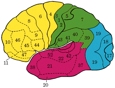
identify Brodmann's areas for cortical areas involved in motor control: Motor control is on the Pre-Central side. That would include areas 4 & 6. See graphic below for reference. Also go to the Brodmann's Area Chart for more info on this topic.
describe the sources of corticospinal tract and patterns of innervation:
discuss the control primary motor cortex has over voluntary movement, in term of specificity, muscle parameters and direction of movement: Primary motor cortex has organized vertical columns. Microstimulation of any area there can result in discrete movements of individual muscles. IE. Stimulation in a vertical column in the hand area may evoke flexion of a digit. Neurons in the same columnar array will receive somatosensory feedback from the patch of skin on the glabrous surface of the digit, which is the area that would come in contact with a surface when the digit is flexed, as to grasp an object. These connections are part of a long-latency reflex circuit. The sensory info reaches the cortex from the ascending sensory systems indirectly via synapses in the thalamus and the primary sensory cortex. The inference here is that motor cortical neurons are informed of the result of their output. There are small cortical neurons that encode movement direction. Some neurons fire rapidly for a movement in one direction but are silent for a movement in the opposite direction.
what is the role of long-loop reflexes: ????????
compare premotor cortex, supplementary motor area and posterior parietal area: The premotor cortex is Brodmann’s area 6. This region contains a somatotopic representation of the body musculature that is complete, although less precisely so than primary motor cortex (MI). The premotor cortex receives input from sensory areas of the parietal cortex and projects to MI, the spinal cord, and the reticular formation. The latter gives rise to reticulospinal fibers, which, in turn, influence spinal motor neurons that innervate paravertebral and proximal limb musculature. It has been suggested that the premotor cortex is involved in the preparation to move. It organizes those postural adjustments that are required to make a movement. The supplementary motor cortex occupies the area of 6 that lies anterior to MI near the convexity and extends onto the medial wall of the hemisphere near the paracentral gyri. It also contains a map of the body musculature, although not as precise as MI. It receives input from the parietal lobe and projects to MI and directly to the reticular formation and cord. Stimulation of this area can evoke movements. In contrast to the single-muscle movements evoked by MI stimulation, however, these movements involve sequences or groups of muscles and orient the body or limbs in space. In addition, higher stimulus intensities are required than for MI, and bilateral movements of the hands or upper extremities are often produced. This area, then, is involved in organizing or planning the sequence of muscle activation required to make a movement, whereas the MI mainly functions to execute the movement. The posterior parietal area comprises area 5 and 7, which occupy the superior parietal lobule. These areas carry out some of the "background computations" necessary for making movements in space. To organize such a movement, it is necessary to collate input from a variety of sensory systems to create a map of space and to compute a trajectory by which a body part can reach its target. Area 5 receives extensive projections from somatosensory cortex and input from the vestibular system, whereas area 7 processes visual info related to the location of objects in space. Both areas project primarily to supplemental and premotor cortices and have few spinal or brainstem targets. This area is responsible for eye-hand coordination and reaching movements.
compare the different regions of the cerebellum in terms of inputs, outputs, and their roles in movement: The cerebellum can be divided into the flocculonodular, medial , vermal, and hemisphere sections. The flocculonodular region receives input from the vestibular nuclei only and has output there as well for balance and eye movement. The vermal region in the very center of the cerebellum receives input from vestibular and visual areas. The output from the vermis goes to vestibular nuclei. The medial region receives input from the spinal and trigeminal nuclei as sensory info and has outputs to the medial parts of the spinal gray to the spinal and trigeminal areas and as you move more lateral to the locomotive system. The hemispheres receive input from the pontine nuclei in the pons, which receives cortical info. The outputs their help to organize preplanned movements to be coordinated and smooth. The output is called the dentate nucleus and goes back to motor and premotor for motor planning.
discuss the mechanisms involved in motor learning: The cerebellum is involved in the learning of a variety of relatively simple reflexive motor behaviors. It is extremely difficult to produce specific modifications in certain reflexes without this structure. These include the adaptation of the vestibulo-ocular reflex and the classic conditioning of reflexes evoked by aversive stimuli, such as the eyeblink and withdrawal reflexes. Lesions of this structure impair the performance of certain previously learned behaviors. This structure also plays a role in the acquisition of several motor behaviors including skilled volitional movements, although the nature of its contribution has not been characterized. The cerebellum’s participation in memory storage for certain reflex behaviors is unclear, although it is not considered to be an essential storage site for engrams related to complex volitional movements.
describe the afferent, internal and efferent connections of the basal ganglia: There are many connections within the basal ganglia. There are afferent connection from the substantia nigra, the cortex, the premotor and supplemental motor areas of motor cortex, primary somatosensory area, the thalamus, the subthalamus, the cerebellum. Internal connections intertwine from one region to the other starting with the caudate, the putamen and the globus pallidus. The efferent connections go to the substantia nigra, the inferior olive, the cerebellum, the motor and premotor cortex and the thalamus.
describe symptoms and pathophysiology of Huntington's disease, Parkinson's disease and tardive dyskinesia: Parkinson’s has the characteristics of akinesia- the impairment of initiation of a movement and bradykinesia- a reduction of velocity and amplitude of movement. Pts with akinesia have a generalized disruption of the role of the basal ganglia in planning and generating programmed movements. Bradykinesia is due to a disruption of the balance btw the outflows of the direct and indirect pathways to the thalamus. The result is an increase in the activation of antagonist muscle. Thus, the observed symptoms are due to an inappropriate activation of the antagonistic muscles, and not necessarily due to an overall decrease in muscular activity. Parkinson’s is correlated pathophysiologically with the loss of the melanin-containing dopaminergic neurons of the nigral complex that project to the striatum. Dopamine has opposite effects on the direct and indirect pathways: It is excitatory to striatal cells feeding into the direct pathway and inhibitory to those of the indirect pathway. The loss of the dopaminergic neurons changes the chemistry of the striatum which favors an increase in the activity of the striatal projections to the lateral pallidum and a decrease in the activity of the striatal projections to the medial pallidum and substantia nigra. Parkinson’s is characteristic of tremor at rest, cogwheel rigidity, akinesia, bradykinesia, disturbances of eye movement and loss of postural reflexes.
Huntington’s disease is characteristic of choreiform movements which are generalized irregular dancelike movements of the limbs. Similar movements may occur in oral and facial muscles. There is an initial selective loss of the medium spiny cells in the striatum, which project to the lateral pallidum, and of acetylcholine-containing neurons in the striatal complex. It is thus likely that the neurons specifically associated with the indirect pathway are lost. This disturbance can most easily be explained by the disruption of the indirect pathway through the motor loop, resulting from the loss of excitatory subthalamopallidal neurons. The balance btw excitation of pallidothalamic neurons and their inhibition is skewed. The result is a decrease in the net amounts of inhibition of thalamic cells, which results in more activity in the cerebral cortex. Huntington’s is a progressive untreatable disorder in which pts lose their ability to function and show increasing dementia and death after 10 –15 years after onset. It is an autosomal dominant genetic disorder. Glutamate excitotoxicity is thought to be the mechanism for the striatal neuron death.
Tardive dyskinesia is a basal ganglia disorder that is iatrogenic in nature, that is, caused by medical intervention for anther disease. This condition is caused by chronic treatment for neuroloeptic medications such as phenothiazines and haldol. The manifestation of this condition is uncontrolled involuntary movements, particularly of the face and tongue and cogwheel rigidity. These symptoms may be temporary or permanent. The action of these neuroleptic drugs is to block dopaminergic transmission throughout the brain. The primary target cells are those in the ventral tegmental area that form the mesolimbic dopaminergic pathway. Prolonged treatment with the drugs leads to a hypersensitivity at the D3 dopamine receptor, which causes an imbalance in the nigrostriatal influence on the basal ganglia motor loop and ultimately results in movement disorders.
Last Updated 04/10/00 12:27:16 PM
Return To The MNA2001 Homepage