readings required or recommended: Guyton and Hall (9th edition). Chap. 38; Ganong (19th edition), Chap. 34
pulmonary blood flow: Passage of blood from the heart to the lungs for purification and the return to the heart. The blood flows from the right cardiac ventricle through to the lungs, there to be oxygenated, then back to the left cardiac atrium. Taber's. Nearly all the blood in the body passes via the pulmonary artery to the pulmonary capillary bed, where it is oxygenated and returned to the left atrium via the pulmonary veins. Ganong, page 619
anatomical shunt: Part of the bronchial blood flow. There are anastomeses between the bronchial capillaries and the pulmonary capillaries and veins, and although some of the bronchial blood enters the bronchial veins, some enters the pulmonary capillaries and veins, bypassing the right ventricle. Also, some blood flows from the coronary arteries into the chambers of the left side of the heart thus bypassing the alveolar capillaries. Ganong page 630
intrapulmonary shunt: When there is a blockage (fluid infiltration, chronic obstruction, acute obstruction, bronchial constriction, pneumonia) in the lung preventing oxygenated air to flow where it normally would to a blood fed alveolar. From Class Notes
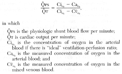
physiological shunt: The total quantitative amount of shunted blood per minute. The greater the physiologic shunt, the greater the amount of blood that fails to be oxygenated as it passes through the lungs. Guyton page 510-511
shunt equation: Guyton page 510
hypoxic vasoconstriction: When a bronchus or a bronchiole is obstructed, hypoxia develops in the under-ventilated alveoli beyond the obstruction. The O2 deficiency apparently acts directly on VSM in the area to produce constriction, shunting blood away from the hypoxic area. Ganong page 632
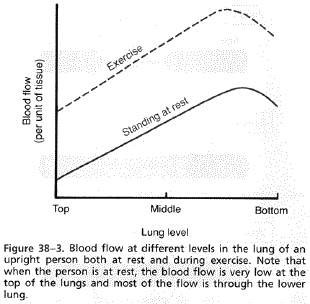
hydrostatic pressure gradients within the lung: In the normal upright adult, the lowest point in the lungs is about 30 cm below the highest point. This represents a 23 mm Hg pressure difference, about 15 mmHg of which are above the heart and 8 below. That is, the pulmonary arterial pressures in the uppermost potion of the lung of a standing person are bout 15 mmHg less than the pulmonary arterial pressure at the level of the heart, and the pressure in the lowest portion of the lungs is about 8 mmHg greater. Such pressure differences have profound effects on blood flow thought the different areas of the lungs. Guyton page 493
zone 1: The very top of the lungs. No blood flow during any part of the cardiac cycle because the local capillary pressure in that area of the lung never rises higher than the alveolar pressure during any part of the cardiac cycle. Guyton page 494
zone 2: The middle to top of the lungs. Intermittent blood flow only during the pulmonary arterial pressure peaks because the systolic pressure is greater than the alveolar pressure but the diastolic pressure is less than alveolar pressure. Guyton page 494
zone 3: The lower portions of the lungs. Continuous blood flow because the alveolar capillary pressure remains greater than alveolar pressure during the entire cardiac cycle. Guyton page 494
zone 4: ??? Area in the very base
orthopnea: Respiratory condition in which there is discomfort in breathing in any but erect sitting or standing position. Taber's
pulmonary artery wedge pressure (PAWP): Pressure measured in the pulmonary artery at its capillary end. Since no valve is present between this location and the left atrium, the measurement reflects left atrial pressure, and, in the presence of a competent mitral valve, the measurement provides an indication of left ventricular end-diastolic pressure. Normally 20-30/8-12 mmHg Taber's
pulmonary interstitial fluid dynamics: Pulmonary capillaries and the pulmonary lymphatic system normally maintain a slight negative pressure in the interstitial spaces, then it is clear that whenever extra fluid appears in the alveoli, it will simply be sucked mechanically into the lung interstitium through the small openings between the alveolar epithelial cells. Then the excess fluid is either carried away through the pulmonary lymphatics or absorbed into the pulmonary capillaries. Thus, under normal conditions, the alveoli are kept in a "dry" state except for a small amount of fluid that seeps from the epithelium onto the lining surfaces of the alveoli to keep them moist. Guyton page 496
describe using words, charts, and/or graphs FLUID BALANCE WITHIN THE LUNG, identifying the factor or factors which normally guard against PULMONARY EDEMA: Pulmonary capillary pressure is about 10 mmHg, whereas the oncotic pressure is 25 mmHg, so that there is an inward-directed pressure gradient of about 15 mmHg which keeps the alveoli free of fluid. Ganong page 630-631
Pulmonary capillaries and the pulmonary lymphatic system normally maintain a slight negative pressure in the interstitial spaces, then it is clear that whenever extra fluid appears in the alveoli, it will simply be sucked mechanically into the lung interstitium through the small openings between the alveolar epithelial cells. Then the excess fluid is either carried away through the pulmonary lymphatics or absorbed into the pulmonary capillaries. Thus, under normal conditions, the alveoli are kept in a "dry" state except for a small amount of fluid that seeps from the epithelium onto the lining surfaces of the alveoli to keep them moist. Guyton page 496
All the factors that tend to prevent edema in the peripheral tissues also tend to prevent edema in the lungs. That is, before positive interstitial fluid pressure can occur and cause edema, all the following factors must be overcome:
Guyton page 497
define the terms ANATOMICAL SHUNT, INTRAPULMONARY SHUNT and PHYSIOLOGICAL
SHUNT, clearly distinguishing among the three:
anatomical shunt: Part of the bronchial blood flow.
There are anastomeses between the bronchial capillaries and the pulmonary
capillaries and veins, and although some of the bronchial blood enters the bronchial
veins, some enters the pulmonary capillaries and veins, bypassing the right
ventricle. Also, some blood flows from the coronary arteries into the
chambers of the left side of the heart thus bypassing the alveolar capillaries.
Ganong page 630
intrapulmonary shunt: When there is a blockage (fluid
infiltration, chronic obstruction, acute obstruction, bronchial constriction,
pneumonia) in the lung preventing oxygenated air to flow where it normally would
to a blood fed alveolar. From Class Notes
physiological shunt: The total quantitative amount of
shunted blood (anatomical shunt + intrapulmonary shunt) per minute. The greater the physiologic shunt, the greater
the amount of blood that fails to be oxygenated as it passes through the
lungs. Guyton page 510-511
describe using graphs and/or mathematical expressions the MEASUREMENT OF SHUNT FLOW, and describe a method of distinguishing the contribution of true intrapulmonary and anatomical shunts from the contribution of regions with a low ventilation-perfusion ratio: Not sure how to distinguish between the two.
Guyton page 510
compare and contrast using words or charts the following CHARACTERISTICS OF THE PULMONARY AND SYSTEMIC CIRCULATIONS: flow rate, volume, compliance, pressures and total resistance:
| Pulmonary | Systemic | |
| Flow Rate | ||
| Volume | ||
| Compliance | Very High | Lower |
| Pressures | ||
| Total Resistance |
describe in words the use of the Swan-Ganz catheter to determine PULMONARY ARTERY WEDGE PRESSURE, and discuss the relationships among pulmonary artery wedge pressure, left atrial pressure, left ventricular end diastolic pressure, and left ventricular end diastolic volume: Achieved by inserting a catheter through the right side of the heart and then through the pulmonary artery into one of the small branches of the pulmonary artery and finally, pushing the catheter until it wedges tightly in the artery. The pressure is then measured through the catheter. Because all blood flow has been stopped in the small artery and the blood vessels extending from the artery make almost direct connection through the pulmonary capillaries with the blood in the pulmonary veins, this wedge pressure is usually only 2 to 3 mmHg greater than the left atrial pressure. Since no valve is present between this location and the left atrium, the measurement reflects left atrial pressure, and, in the presence of a competent mitral valve, the measurement provides an indication of left ventricular end-diastolic pressure. Left ventricular end diastolic volume??? Guyton page 492 & Taber's
describe in words, charts and/or graphs the REGULATORY MECHANISMS CONTROLLING PULMONARY BLOOD FLOW and its regional distribution within the upright lung, including HYDROSTATIC PRESSURE GRADIENTS, the phenomenon of HYPOXIC PULMONARY VASOCONSTRICTION, and the EFFECTS OF AUTONOMIC INNERVATION:
Guyton page 493
Gravity has a relatively marked effect on the pulmonary circulation. In the upright position, the upper portions of the lungs are well above the level of the heart, and the bases are at or below it. Consequently, there is a relatively marked pressure gradient in the pulmonary arteries from the top to the bottom of the lungs, because of the effect of gravity, and a resulting linear increase in pulmonary blood flow (See graph below) from the apices to the bases of the lungs. In the normal upright adult, the lowest point in the lungs is about 30 cm below the highest point. This represents a 23 mm Hg pressure difference, about 15 mmHg of which are above the heart and 8 below. That is, the pulmonary arterial pressures in the uppermost potion of the lung of a standing person are bout 15 mmHg less than the pulmonary arterial pressure at the level of the heart, and the pressure in the lowest portion of the lungs is about 8 mmHg greater. Such pressure differences have profound effects on blood flow thought the different areas of the lungs. Guyton page 493
The lungs can be separated into 3 zones of blood flow.
When a bronchus or a bronchiole is obstructed, hypoxia develops in the under-ventilated alveoli beyond the obstruction. The O2 deficiency apparently acts directly on VSM in the area to produce constriction (hypoxic vasoconstriction), shunting blood away from the hypoxic area and putting to another more oxygenated area of the lungs. Ganong page 632
Although nerves innervate the lung tissues, it is doubtful that they have a major function in normal control of pulmonary blood flow. Normally, stimulation of the vagal fibers to the lungs causes a slight decrease in pulmonary vascular resistance, and stimulation of the sympathetics causes a slight increase in resistance, both effects perhaps too little to be more than marginally important. Guyton page 493
describe the effect of pulmonary edema on the ability of oxygen and carbon dioxide to diffuse across the respiratory membrane (pulmonary diffusion capacity): I can't find this in the books or notes.
Last Updated 04/10/00 12:27:08 PM
Return To The MNA 2001 Homepage