circulatory failure: Generalized inadequacy of blood flow throughout the body to the extent that the body tissues are damaged because of too little flow, especially too little delivery of oxygen and other nutrients to the tissues cells.
cardiac failure: Failure of the heart to pump enough blood to satisfy the needs of the body.
shock: A result of inadequate cardiac output or circulating blood volume to support the body.
low-output failure: Most often occurs after acute MI’s or after a prolonged period of progressive cardiac deterioration. The heart becomes incapable of pumping even the minimal amount of blood flow required to keep the body alive. Consequently, all the body tissues begin to suffer and even deteriorate, often leading to death within a few hours to days.
high-output failure: Signs of cardiac failure because the left or right atrial pressures are much too high and there usually is an accumulation of edema. The problem is an overloading of the heart with too much venous return. It could be caused by an AV fistula, beriberi or thyrotoxicosis.
ventricular function curve: Guyton p 241
fatigue: A state of increased discomfort and decreased efficiency resulting from prolonged exertion.
Edema: An abnormal accumulation of fluid in the intercellular spaces of the body. In the cardiac system edema is the manifestation of congestive heart failure due to increased venous and capillary pressures and often associated with renal sodium retention.
Preload: Preload is the tension applied to the walls of the heart as it stretches during diastole (ventricular filling). The volume of blood is translated to the actual tension and preload is the stretch applied to the heart before it starts to contract so it is associated with filling. You can indirectly measure preload by measuring filling pressures and end diastolic volume (EDV). An increase in preload is usually by increasing EDV which is usually caused by an increase in fluid or in filling pressures. The increased pressure will increase the volume, which will increase the wall tension and thus increases contractile force. In heart failure there is often fluid retention and therefore increased vascular pressures which increases filling pressures so that you have a larger than normal EDV which increases preload. A stiff heart due to HTN will require higher than normal EDV to fill the heart so the filling pressures are higher.
afterload: The wall tension on the heart in order to eject the blood is the afterload. If there is an increase in arterial pressure then ventricular pressure must also increase. The wall tension in the heart will have to increase to overcome the art. pressure. If someone has a stenotic aortic valve there is high resistance and the vent pres must increase to eject blood. The wall tension must increase to generate enough pressure. Afterload then, is increased by an increase in arterial pressure and by valvular pathology, also by HTN and HOC. In aortic stenosis you often have low arterial pressure as a compensatory mechanism to reduce the afterload and preserve the heart function.
myocardial contractility: This is the ability of the heart to increase its strength of contraction. It can do it in two ways. The first is by an increase of Ca++ ions infused into the myocardial cells. By increasing the [Ca++] the number of actin/myosin crossbridges are exposed leading to a stronger, more forceful contraction. There will also be a greater emptying of the ventricle as a result which will also increase the stroke volume. This is the inherent inotropy of the heart. The 2nd way to increase contractility is to use Starling’s Law and increase preload. Preload will give you a larger volume in the ventricle, which will passively stretch the walls of the ventricle by applying tension to the walls. Look at the length tension curve and see that as you increase the passive stretch of the walls you will increase the force of contraction.
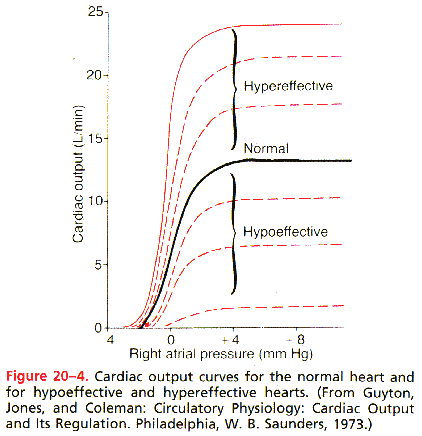
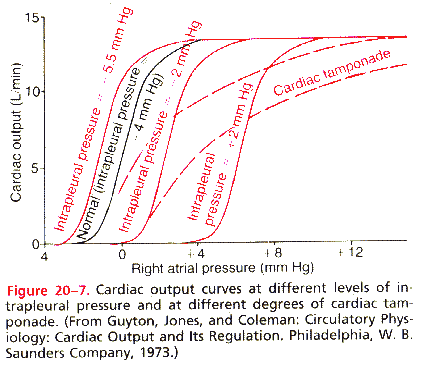
cardiac output curve: Guyton p 244
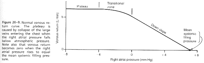
venous pressure curve: Guyton p 245
Surface Tension Law: ???? Law of LaPlace says tension is equal to the pressure x radius divided by wall thickness.
Frank-Starling mechanism: In Starling’s Law the level of contractility is based on the changes in fiber length. Preload is the stretch on the wall or the tension applied on the wall just before contraction. This tension works on the passive stretching of the muscle before contraction. You can increase the tension by increasing the volume or the pressure and this will increase the muscular contraction and thus increase stroke volume.
myocardial hypertrophy: The structural remodeling that occurs from chronic changes in pressure or tension in the wall of the ventricle resulting in a larger than normal heart with wall thickening of the ventricle. In concentric hypertrophy the remodeling is a result of a chronic increase in afterload due to an increase in MSAP. The ratio between wall thickness and pressure will normalize the wall tension. It will reduce the tension per square surface area since there is more surface area to spread out the tension thus normalizing the tension. The radius often decreases as a result. Eccentric hypertrophy is due to a chronic increase in preload. There is a stronger than normal force of contraction since the filling pressures are greater and so the SV is greater. Starling’s Law states that the greater the tension or stretch the greater the contraction. Therefore wall tension is chronically increased and this results in remodeling of the ventricular wall again but this time the CXR shape is elongated and off center. This thickness is also associated with an increase in radius to keep their ratio equal.
myocardial energetics: The consumption of oxygen during the cardiac cycle. ATP utilization is at its highest during systole.
coronary blood flow: Occurs during diastole in the middle to late phases and then is squeezed off during the onset of systole and into the end of the isovolumetric relaxation phase.
pulmonary edema: An accumulation of fluid in the interstitial spaces within the lung thus forming a barrier between capillary flow and the alveoli which results in the impairment of gas exchange. Pulmonary edema can be set off by left ventricular failure when blood begins to back up into the lungs due to the failed LV. This increase in blood elevates the pulm capillary pressure and the fluid begins to leak out into the interstitium. This will decrease the oxygenation of the blood which further weakens the heart and causes peripheral vasodilatation which causes an increase in the venous return and further increases the back up of blood in the lungs.
systemic edema: This is caused by right sided heart failure. The fluid retention actually occurs because of the kidneys. This retention increases the mean systemic filling pressures which results in increased tendency for blood to return to the heart which elevates the right atrial pressure back toward normal. The capillary pressure also rises markedly thus causing loss of fluid into the tissues and development of severe edema. The decreased cardiac output due to the failure will decrease the GFR, which will decrease u/o. This will activate the RAA system, which will cause more vasoconstriction and increased reabsorption of water and salt into the renal tubules. Aldosterone also causes reabsorption of sodium, which increases the osmotic concentration of the ECF everywhere in the body, which stimulates ADH hormone secretion which promotes further tubular reabsorption of water.
hemorrhagic shock: This shock decreases the filling pressure of the circulation, which decreases venous return. Cardiac output falls below normal and shock ensues.
neurogenic shock: Shock resulting in a sudden loss of vasomotor tone throughout the body, causing massive dilatation of the veins. The vascular capacity increases so much that even the normal amt of blood becomes incapable of adequately filling the circulatory system. Deep general anesthesia, which could depress the vasomotor center enough to cause vasomotor collapse, spinal anesthesia, which can block the sympathetic outflow from the nervous system, and brain damage can all cause this type of shock. Vasovagal syncope from emotional stimulus can result in sudden loss of vasomotor tone.
cardiogenic shock: Shock when the cardiovascular system fails due to inadequate cardiac pumping. The survival rate even with therapy is only 15%.
septic shock: Widely disseminated bacterial infection to many areas of the body, with the infection being borne through the blood from one tissue to another and causing extensive damage. This type of shock occurs most often other than cardiogenic shock in hospitals today. It is often associated with a high fever, marked vasodilation throughout the body, especially in the infected tissues, high cardiac output, sludging of the blood, presumably caused by red cell agglutination in response to degenerating tissues, and development of microclots in widespread areas of the body, a condition called DIC which also causes the clotting factors to be used up so that hemorrhages occur into many other tissues, especially the gut wall and the intestinal tract.
anaphylactic shock: An allergic shock condition in which cardiac output and arterial pressure often fall drastically as a result of the antigen-antibody reaction that takes place immediately after an antigen to which the person is sensitive has entered the circulation. The basophils of the blood and mast cells in the pericapillary tissues release histamine or a histamine-like substance which causes an increase in vascular capacity because of venous dilatation and dilatation of the arterioles with resultant greatly reduced arterial pressure, and greatly increased capillary permeability with rapid loss of fluid and protein into the tissue spaces.
non-progressive shock: Also called the compensated stage of shock, the normal circulatory compensatory mechanisms will eventually cause full recovery without help from outside therapy. The negative feedback controls kick in to control the shock. The baroreceptors, central nervous system ischemic response are elicited when the arterial pressure falls; the reverse stress-relaxation of the circulatory system causes the blood vessels to contract down around the diminished blood volume, so that the blood volume that is available will more adequately fill the circulation. The formation of angiotensin constricts the peripheral arteries and causes increased conservation of water and salt in the kidneys. The formation of vasopressin (ADH) will constrict the peripheral arteries and veins and greatly increase water retention by the kidneys. There will also be a compensatory mechanism to return the blood volume back toward normal by absorption of large quantities of fluid from the intestinal tract, the interstitial spaces of the body and conservation of water and salt by the kidneys, and increased thirst and appetite for salt.
progressive shock: The shock becomes steadily worse until death. This is a result of positive feedback that causes a viscious circle of progressively decreasing cardiac output. The vasomotor center will utilize all the factors mentioned above but as the blood flow to the vasomotor center further diminishes the output from this area will steadily slow down until it fails altogether. There will be sluggish blood flow in the microvessels, which will cause blockages in them, which will further progress the shock. There is an increased capillary permeability after many hours of capillary hypoxia, which further decreases the blood volume. The ischemic tissues release endotoxins, which causes greatly increased cellular metabolism and also causes cardiac depression.
irreversible shock: The shock has progressed to such an extent that all forms of known therapy are inadequate to save the person’s life, even though, for the moment, the person is still alive. Signs of generalized cellular deterioration occur throughout the body. The liver especially is affected due to the lack of nutrients to support the normal rate of metabolism and to the extreme vascular exposure of the liver cells to any toxic or other abnormal metabolic factor in shock.
traumatic shock: Often the shock results from hemorrhage caused by the trauma but it can also occur even without hemorrhage because contusion of the body can damage the capillaries sufficiently to allow excessive loss of plasma into the tissues. This results in greatly reduced plasma volume with resultant hypovolemic shock. The pain associated with serious trauma can also be an additional aggravating factor because pain sometimes seems to inhibit the vasomotor center, thereby inhibiting sympathetic signals to the circulation which increases the vascular capacitance and reduces the venous return.
describe in words or graphs how GENERALIZED CIRCULATORY FAILURE can arise from failure of 1) the cardiac pump or 2) the peripheral circulation: Failure of the cardiac pump can cause a spiraling downward into the blackhole. The failure results in a decrease in cardiac output, which further decreases the perfusion of the heart, and a further deterioration in overall heart function.
Causes for the cardiac pump to fail are due to:
CIRC. FAILURE arises from the peripheral circulation due to:
list and describe possible CAUSES OF CARDIAC FAILURE, and describe the pathophysiological bases for two of the major CONSEQUENCES OF HEART FAILURE, fatigue and increased central venous pressure: Arises when demands placed on the heart exceed it's capacity to work, initially with exercise, and eventually even at rest. Compensatory mechanisms of stretching (Starling's law) and increased contractility no longer match demands. Fatigue occurs due to sub-optimal blood flow to tissues. Increased central venous pressure occurs because cardiac output falls and blood "backs up".
describe in words and diagrams the sequence of ACUTE and CHRONIC PHYSIOLOGICAL COMPENSATORY MECHANISMS that follow a moderately severe myocardial infarction: Acutely, ischemic heart causes pain (& possibly decreased CO) which stimulates the SNS. SNS causes increase in HR, contractility, TPR, and decrease in compliance. Overtime, Na+/H20 retention develop to increase cardiac output via starling's law. If SNS stimulation is sustained, remodeling of the heart occurs, with down regulation of the receptors.
compare and contrast the ADVANTAGES AND DISADVANTAGES of increased PRELOAD (signified by EDEMA), increased SYMPATHETIC STIMULATION (signified by TACHYCARDIA), and MYOCARDIAL HYPERTROPHY (indicated by the LEFT AXIS DEVIATION) as compensatory mechanisms in heart failure
|
ADVANTAGES |
DISADVANTAGES |
|
|
increased PRELOAD |
The renal attempt to compensate for the failure will be to increase the blood volume which will increase the SV due to a better stretch with Starling’s Law |
An increase in MVO2 Edema both peripheral and pulmonary depending on the side of heart failure. |
|
increased SYMPATHETIC STIMULATION |
This will increase the contractility, heart rate, TPR which will increase the MSAP and a decrease in venous compliance which will increase CVP and preload |
Increase MVO2 and down-regulation |
|
MYOCARDIAL HYPERTROPHY |
Increase # of sarcomeres, increase in contractile force, normalized wall tension |
Increase in mass, increase in flow requirements, decrease contractility |
describe in words circumstances that might lead to DECOMPENSATED HEART FAILURE and describe the physiological rationale for the clinical treatment of the decompensation; Hypertropic myocardium doesn't contract well, and volume effect (starling's law) cannot be further magnified.
"Dig & Diuretics" --- increase the contractility and lower volume to get the stretch back into a productive zone.
Earlier on:
Ace inhibitors and Beta blockers stop remodeling process which leads to
pathological hypertrophy, as well as lowering O2 demand (beta block decreases
contractility, ace inhibitor lower afterload).
compare and contrast using words or diagrams the phenomena of SYSTEMIC and PULMONARY EDEMA, with respect to the PATHOPHYSIOLOGY underlying each and their respiratory and cardio-vascular CONSEQUENCES: Backward verses forward failure. In Backward failure, CO drops, and blood backs up into the lungs. Pulmonary edema develops and central venous pressues go up. Can be due to systolic failure such as in aortic stenosis, or diasystolic failure such as ishemic tissue with loss of compliance. In forward failure, have insufficient pressure gradient to perfuse kidneys and therefore fluid collects peripherally. There is a much great margin for volume increase within the systemic circulation due to high venous compliance.
define CIRCULATORY SHOCK, and describe in words and diagrams its major stages, pathogenesis, and its effect on cardiac output and mean systemic arterial pressure: Circulatory shock is generalized inadequacy of blood flow throughout the body to the extent that the body tissues are damaged because of too little flow, especially too little delivery of oxygen and other nutrients to the tissue cells. It can be caused by any factor that reduces CO. There are two factors that reduce the CO: cardiac abnormalities that decrease the ability of the heart to pump blood and factors that decrease the venous return. In most cases the MSAP will be decreased but in can be increased from the powerful nervous system reflexes we have.
using HEMORRHAGIC SHOCK as a model, discuss in words and using graphs or charts each of the following:
define each of the following and describe possible causes and probable
effects: NEUROGENIC SHOCK, TRAUMATIC SHOCK, ANAPHYLACTIC SHOCK:
neurogenic shock: Shock resulting in a sudden loss of vasomotor tone
throughout the body, causing massive dilatation of the veins. The vascular
capacity increases so much that even the normal amt of blood becomes incapable
of adequately filling the circulatory system. Deep general anesthesia, which
could depress the vasomotor center enough to cause vasomotor collapse, spinal
anesthesia, which can block the sympathetic outflow from the nervous system, and
brain damage can all cause this type of shock. Vasovagal syncope from emotional
stimulus can result in sudden loss of vasomotor tone.
traumatic shock: Often the shock results from hemorrhage caused by
the trauma but it can also occur even without hemorrhage because contusion of
the body can damage the capillaries sufficiently to allow excessive loss of
plasma into the tissues. This results in greatly reduced plasma volume with
resultant hypovolemic shock. The pain associated with serious trauma can also be
an additional aggravating factor because pain sometimes seems to inhibit the
vasomotor center, thereby inhibiting sympathetic signals to the circulation
which increases the vascular capacitance and reduces the venous return.
anaphylactic shock: An allergic shock condition in which cardiac
output and arterial pressure often fall drastically as a result of the
antigen-antibody reaction that takes place immediately after an antigen to which
the person is sensitive has entered the circulation. The basophils of the blood
and mast cells in the pericapillary tissues release histamine or a
histamine-like substance which causes an increase in vascular capacity because
of venous dilatation and dilatation of the arterioles with resultant greatly
reduced arterial pressure, and greatly increased capillary permeability with
rapid loss of fluid and protein into the tissue spaces.