Na+/K+ ATPase: This is the active transport process that pumps sodium ions outward through the cell membrane and at the same time pumps potassium ions from the outside to the inside. The pump is responsible for maintaining the Na+ and K+ concentration differences across the cell membrane as well as for establishing a negative electrical potential inside the cell.
Fast Na+ channels: At onset of the action potential the sodium channels instantaneously become activated and allow a huge increase in conductance to sodium. This allows a huge inflow of sodium ions making the interior of the cell much more positive and beginning the action potential. These fast Na+ channels are located in atrial and ventricular muscle and Purkinje fibers and not in the SA or AV node. The fast inflow of sodium is responsible for the rapid spike on the action potential diagram.
Slow Na+/Ca++ channels: The slower Na+/Ca++ channels are located in all
areas of the heart. In the SA node there is a tendency for sodium to leak into
the cells, this tends to keep the resting membrane potential at a more positive
# closer to –55. Around –60 the fast Na+ channels become inactivated thus
the action potential must rely on the slower Na+/Ca++ channels for the action
potential. As a consequence the rate of rise of the AP is slower and these
channels stay open longer than the fast ones thus allowing the inflow of Ca++
and Na+ ions. The Ca++ ions are also used by the cells to activate cross-bridge
formation and thus cause a contraction of the muscle.
K+ channels: After 100-150 m/sec the slow Na+/Ca++ channels close and the K+ channels open allowing large quantities of K+ to leave the cell which terminates the AP. The K+ channels remain open a few tenths of a second longer and continue to allow K+ to leave the cell. This causes an excess negativity in the cell or hyperpolarization. This hyperpolarization is not maintained due to the sodium leak channels which can now continue and allow a slow leak to of sodium in which raises the membrane potential and once it reaches –55 the slow channels open and set off another AP. This whole inflow of Na+ ions gives the SA node its automaticity.
Activation: Refers to the resting membrane potential reaching –55 and suddenly the AP gets set off.
Inactivation: Refers to the blocking of the fast Na+ channels in the SA node which could possibly be reactivated after defibrillation for treatment of Vfib. The fast Na+ channels technically could be reset for the first beat after the defib but would then revert back to being blocked as the resting membrane potential slowly rises up due to the leak in of sodium through the sodium leak channels and since the remaining resting potentials will not go below –60, the fast Na+ channels will be inactive again.
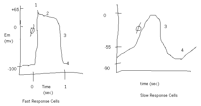
Phase 0 of the cardiac action potential: Phase 0 in the SA and AV node is the period of increased conductance (g) to the Ca++ (L-type) slow channels which allow the slower magnitude and rate of rise of the AP. Phase 0 in the fast response cells is the rapid opening of the fast Na+ channels, which allow Na+ entry due to an increase g to Na+.
Phase 1: Slow response has no phase 1. Fast response: this is the increased g to K+ (transient increase of g to the K channel).
Phase 2: Also no 2 in the slow response. Fast response, this is the plateau phase due to the slow Ca++ channels (L-type) that are open longer and allow an increase g to Na+ and to Ca++ and a decreased g to K+. There is a large equilibrium gradient for K+ at this point so there is a large electrical gradient for K+.
Phase 3: Slow response: this is where there is a decrease g to Ca++ and an increased g to K+ - slight delay of K+ opening then they do open and net movement of cations is out. The Fast response cells will have an increase in g for K+ and a decrease in g for Ca++ - open K+ channels are delayed which causes repolarization.
Phase 4: Slow response: K+ channels close and slowly Na+ leak overwhelms the K+ outflow and slowly increases the membrane potential. There is an increase in g to Ca++ to the T-type channels which gives the kick to the last segment of depolarization to send it over threshold then the L-type channels open and you have phase 0. Fast response: The quiet resting phase since there normally isn’t any leak of sodium in these cells to cause automaticity unless the cells become hypoxic or are stimulated and could then spontaneously depolarize.
Depolarization: The part of the AP when the membrane becomes permeable to positively charged ions that want to satisfy their concentration gradient and come into the cell thus causing the cell to become rapidly more positive inside.
Plateau: The prolonged period in the depolarization phase of the cardiac muscle.
Repolarization: The rapid diffusion of the potassium ions to the exterior of the cell re-establishes the normal negative resting membrane potential.
Absolute refractory period: The period of time during the AP that the cell can not be restimulated to cause another AP.
Relative refractory period: The period of time in the last m/sec of the AP when a strong enough stimulus could set off another AP but the amplitude of this next AP would most likely be considerably less.
Spontaneous Phase 4 depolarization: As discussed above, refers mostly to the Slow response cells although in the right situation occur in the Fast response cells – drugs, hypoxia, and damage.
Autorhythmicity: The inherent nature of the different cells of the myocardium to provide a lifetime of AP’s and a backup plan should one section or another fail. SA node beats at a rate of 60-70, AV node – 40-60, Purkinje fibers 30 and ventricular muscle 20-30.
Nodal potentials: ? As above.
Conduction velocity: Refers to the speed in which the electrical impulse is transmitted through the various sections of the heart. SA node to the AV node lasts .12 - .20 sec with the delay being in the AV node to allow the atria to contract and give "atrial kick", the last bit of emptying into the ventricles. The path from the AV node down the bundle of His, to the Purkinje fibers and through the ventricular muscle lasts around .08 sec.
Excitability: The capability of some of the cardiac cells to self-excitation, a process that can cause automatic rhythmical discharge and contraction.
Sino-atrial node (SA): Located in the superior lateral wall of the right atrium immediately below and slightly lateral to the opening of the superior vena cava. The fibers of the node have almost no contractile filaments.
Atrioventricular node (AV): Located in the posterior septal wall of the right atrium immediately behind the tricuspid valve and adjacent to the opening of the coronary sinus.
Atrial conduction pathways: ends of the SA nodal fibers fuse with the surrounding atrial muscle fibers and AP’s originating in the SA node travel outward into these fibers spreading evenly through the entire atrial muscle mass. There are several small bundles of atrial muscle fibers through which the conduction is transmitted. One is the anterior interatrial band and passes through the anterior wall of the atria to the left atrium. There are also the anterior, middle and posterior internodal pathways, which conduct the impulse through the remainder of the atria and end up in the AV node.
Bundle of His: This is the lowest end of the AV node through which the AP travels before it branches into the right and left bundles.
Right/left bundle branches: These lie beneath the endocardium of the two sides of the septum. Each branch spreads downward to the apex of the ventricle, progressively dividing into smaller branches that course around each ventricular chamber and back toward the base of the heart.
Purkinje fibers: These fibers penetrate about one third of the way into the muscle mass and then become continuous with the cardiac muscle fibers.
Ventricular muscle: The cardiac muscle wraps around the heart in a double spiral with fibrous septa between the spiraling layers; therefore, the cardiac impulse does not necessarily travel directly outward toward the surface of the heart but instead angulates toward the surface along the directions of the spirals.
Vector cardiography: The vector of current flow through the heart changes rapidly as the impulse spreads through the myocardium. Vectorcardiogram depicts the changes of in the vectors at the different times during the cardiac cycle.
Electrocardiograph: ??? Does this refer to the picture of the first ECG by Waller in 1887?
Electrocardiogram: I think we all know this one!
Explain the ionic basis for the resting membrane potentials and action potentials found in slow response (nodal) and fast response (Purkinje fibers and myocardium) cells. The slow response cells have sodium leak channels that allow a constant leak of sodium into the cell. This keeps the resting membrane potential more positive, around –55. The constant leak of Na+ into the cell eventually brings the potential up to threshold, which sets off the action potential. The resting membrane potential is too high to keep the fast Na+ channels activated so they become inactivated and the action potential must rely on the slow Na+/Ca++ channels which will become activated at threshold thus giving the look of a slower rate of rise for the AP. These slow channels stay open longer thus giving a plateau phase to the myocardial cell. This allows the cardiac contraction to last longer. K+channels then open as the slow Na+/Ca++ channels close and this causes the repolarization phase. The fast response cells have a resting membrane potential of –90 or so and they do have the fast Na+ channels which when activated cause a rapid inflow of Na+, which gives the very sharp upward spike of the AP. The slow Na+/Ca++ channels then open and give the plateau phase. K+ channels finally open as the other ones close and this is the repolarization phase.
Describe the shape, magnitude, and duration of the Action Potentials from each of the following regions of the heart:
SA node: See the diagram of slow response cells. Duration is <.03 sec
Atrial muscle: See the diagram of the fast response cells. Duration through here is .03 sec.
AV node: See the diagram above of slow response cells. Duration slows through here to allow time for atrial contraction. Duration is around .13 sec.
Purkinje fibers: See fast response cells. Duration here is very fast – 150 times faster than through the AV node about .03 sec.
Ventricular muscle: Also fast cells with a very fast duration of time – around .3-.5 m/sec
Describe, using words and diagrams, the relationship between the sequential depolarization and repolarization of the various portions of the heart during the cardiac cycle and the typical form of the Lead II ECG obtained using electrodes on the body surface. P wave corresponds with the electrical impulse of the action potential of the atria. The PR interval denotes the total time for the impulse to travel to and through the AV node. The QRS denotes the ventricular action potential and subsequent contraction and ST segment is the plateau phase. The T wave is the repolarization of the ventricles. Normal Lead II is with the negative terminal on the right arm, the positive terminal on the left leg and the ground is usually on the left arm. When the right arm is negative with respect to the left leg, the ECG records positively thus the normal tracing should have the PQRST all upright.
List and discuss the ionic basis for AUTOMATICITY in the cardiac conduction system. The SA node is the "pacemaker" of the heart. Its inherent beating rhythm is 60-100bpm. The presence of the leaky sodium channels allow a continuous flow of sodium in through these channels which raises the normal resting membrane potential of the SA node (and the AV node). These channels allow the sodium to leak in to the cell until it reaches threshold around –40 which then sets off the AP.
List the various AUTONOMIC RECEPTOR types found on cardiac muscle and describe the SECOND MESSENGER SYSTEMS associated with each of these receptor types: The SNS has post-ganglionic fibers at the myocardial cells and release norepinephrine (NE). NE binds to the receptors on the heart. These are subtype receptors called beta1 receptors. Binding here of the NE are linked to the 2nd messenger system adenylate cyclase via a stimulatory G-protein. Binding will stimulate the g-protein, which turns on adenylate cyclase, which promotes formation of cyclic AMP. cAMP in the cell causes many functions of myocardial cells. There are cAMP Ca++ channels (L-type) which will be open when cAMP concentrations are high and Ca++ can enter the cell faster. There are also cAMP associated features of the Ca++/ATPase pump which in the presence of high [cAMP] will pump the Ca++ back to the SR faster so will have a stronger contraction with more Ca++ present and the velocity of contraction will be faster. Since the rate of rise of the AP will be higher and the higher the magnitude, the faster the Ca++ will be taken out of the cell and the shorter the AP and the contraction. PSNS has muscarinic receptors on the myocardium, which are linked to the inhibitory G-protein. When stimulated they will decrease adenylate cyclase, which will decrease cAMP, which will have inhibitory effects on the heart.
Describe the mechanisms of action by which sympathetic and parasympathetic stimulation produce CHANGES IN HEART RATE and MYOCARDIAL CONTRACTILIY: The SNS will cause an increase in heart rate, conduction velocity, excitability, and contractility by the above mentioned methods. Also of note is that the basal rate of entry is affected in that there is an increase in the leaky sodium current so the membrane doesn’t go as negative and will have a slightly faster rate of rise. This is the excitability component. Contractility is affected by more Ca++ entering thus more cross-bridges can form. PSNS will cause the opposite as the two have reciprocal innervation of the heart. The nodal cells will have a slower rate of rise and less Ca++ entering and the AP magnitude doesn’t go as high. When the K+ channels are open in the stimulation of the PNS, the resting membrane will go lower since K+ channels will be open longer, which will give the cells a lower hyperpolarization. This also creates more leaky sodium and potassium to move which will take longer to return to threshold and therefore give a slower rate of rise. This slows the HR and lengthens the time between spontaneous impulses. Conduction velocity decreases. The slowest conduction is through the AV node so with PSNS stimulation the speed could be slowed to standstill and give 2nd degree heart blocks. Excitability decreases because the hyperpolarization is more. There will also be decreased contractility of the atria since the ventricles have very little PNS receptors. This would decrease the atrial kick.
Distinguish clearly between the terms "intrinsic rate of rhythmicity" and "rate of action potential conduction": The rate of rhythmicity is the rate at which the SA node fires and is 60-70bpm. The action potential conduction refers to the speed of the actual action potential.
Identify the ANATOMICAL LOCATION and FUNCTIONAL SIGNIFICANCE of the SA node, the AV node, the internodal pathways, the AV bundle, and the right and left bundle branches: See the key words for location and the significance has also been discussed ad nauseum!
Identify the INTRINSIC RATES OF RHYTHMICITY of the SA node, AV node, Purkinje fibers, and ventricular muscle, and identify the normal locus of the cardiac pacemaker. Also discussed already!