Guyton, Chp 9
Sarcolemma: Cell membrane of the muscle fiber. Consists of true cell membrane, called the plasma membrane, and an outer coat made up of a thin layer of polysaccharide material that contains numerous thin collagen fibrils. (Guyton, p. 73)
Sarcomere: Portion of a myofibril (or of the whole muscle fiber) that lies between two successive Z discs. (Guyton, p. 74)
Length-tension relationship: When muscle is at its normal resting length, it contracts with maximum force of contraction. If the muscle is stretched to much greater than normal length before contraction, a large amount of resting-tension develops in the muscle even before contraction takes place; this tension results from the elastic forces of the connective tissue, sarcolemma, blood vessels, nerves, and so forth. The increase in tension during contraction, called active-tension, decreases as the muscle is stretched much beyond its normal length. (Guyton, p.79)
Intercalated discs: Cell membranes that separate individual cardiac muscle cells from one another. Electrical resistance through the intracalated disc is only 1/400 the resistance through the outside membrane of the cardiac muscle fiber because the cell membranes fuse with one another in such a way that they form permeable gap junctions that allow relatively free distribution of ions. (Guyton, p. 107)
Transverse tubular system: Action potential of the cardiac muscle fiber is spread along the transverse (T) tubules. This causes the longitudinal sarcoplasmic tubules to release calcium ions into the sarcoplasmic reticulum. The calcium ions catalyze the chemical reactions that promote the sliding of the actin and myosin filaments along one another to cause muscle contraction. (This present also in skeletal muscle). There is an additional means of calcium entry into the sarcoplasm unique to cardiac muscle. The T tubules of cardiac muscles have 5 times as great a diameter as in skeletal muscle, and the volume of these T tubules is 25 times as great. They contain large amounts of calcium that are released during the action potential. The T tubules also open directly into the ECF in cardiac muscle, so their calcium content depends on the extracellular calcium concentration. At the end of the plateau of the AP, the influx of calcium ions into muscle fiber abruptly stops and calcium is pumped back into the SR and T tubules. Contraction ends. (Guyton, p. 109-110)
Excitation-contraction coupling: The mechanism by which the action potential causes the myofibrils of the muscle to contract. (See Transverse-tubular system). (Guyton, p. 109)
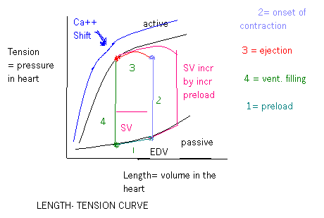
Preload: The degree of tension on the cardiac muscle when it begins to contract. For cardia contraction, is usually considered to be the end-diastolic pressure when the ventricle has become filled. (Guyton, p. 115)
Starling’s Law: The greater the heart muscle is stretched during filling, the greater will be the force of contraction and the greater will be the quantity of blood pumped into the aorta. (ie: within physiological limits). (Guyton, p. 115)
Afterload: The load against which the heart muscle exerts its contractile force. This is the pressure in the artery leading from the ventricle. This correspond to the systolic pressure described by the Phase III curve. (Fig. 9-7, p. 114, Guyton)
Contractility: The ability to shorten or increase tension, applied to muscle. (Melloni’s Illus. Med.Dictionary, p. 110)
dP/dt: Rate of change of the ventricular pressure with respect to time. The rate of rise of ventricular pressure, the dP/dt, in general correlates well with the strength of contraction of the ventricle. (Guyton, p. 118)
End-diastolic volume: The volume filling of the ventricles during diastole (110-112 ml). (Guyton, p. 112)
End-systolic volume: The remaining volume left in the ventricles after systole (40-50 ml). (Guyton, p. 112)
Stroke volume: The volume ejected during systole (about 70 ml). (Guyton, p. 112)
Ejection fraction: The fraction of the end-diastolic volume that is ejected during systole (about 60%). (Guyton, p. 112)
Tricuspid valve: The A-V valve which prevents backflow of blood from the right ventricle to the right atria during systole. (Guyton, p, 112)
Mitral valve: The A-V valve which prevents backflow of blood from the left ventricle to the left atria during systole. (Guyton,p. 112)
Chordae tendineae: Tendinous strands extending from the papillary muscles to the leaflets of the A-V valves of the heart (Melloni’s, p. 98) (Guyton, p. 113)
Echocardiogram: Graphic display obtained from the application of ultrasonic procedures. Used for determining the movement patterns of the heart and its valves, chamber size, wall thickness, and the presence of pericardial fluid. (Melloni’s, p. 140)
Semi-lunar valves: They prevent backflow from the aorta and pulmonary arteries into the ventricle during diastole (aortic and pulmonary valves). (Guyton, p. 112)
Pericardium: The thin, double layered, membranous sac that encloses the heart; the layers are separated by a small amount of fluid which lubricates the constantly rubbing surfaces; the layers fuse as they attach to the great vessels and diaphragm. (Melloni’s, p. 365)
Heart sounds: The vibration of the vanes of the valves and the surrounding fluids under the influence of the sudden pressure differentials that develop, giving off a sound that travels in all directions through the chest. (Guyton, p. 113)
Murmurs: A relatively prolonged series of auditory vibrations resulting from turbulent blood flow. (Melloni’s, p. 280)
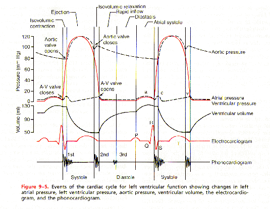
Isovolumic (isovolumetric) contraction: During the period when ventricular contraction has begun, the ventricular pressure abruptly rises, to build up sufficient pressure to open the semilunar valves against the pressures in the aorta and pulmonary artery. Contraction is occurring but no but there is no emptying. Tension is increasing in the muscle but no shortening of the muscle fibers is occuring. (Not strictly true-there is apex to base shortening and circumferential elongation.) (Guyton, p. 112)
Ejection (rapid and reduced phase): When the left ventricular pressure rises slightly above 80 mm Hg and the right slightly above 8 mm Hg, the ventricular pressures nowpush the semilunar valves open. Blood begins to pour out of the ventricles with about 70% of the emptying occurring during the first third of the period of ejection (period of rapid ejection) and the remaining 30% during the next two thirds (period of slow ejection). (Guyton, p. 112)
Isovolumic (isovolumetric) relaxation: At the end of systole when the intraventricular pressures are falling rapidly back to their low diastolic levels and the aortic and pulmonary valves close. The ventricular muscle continues to relax. The ventricular volume does not change. (Guyton, p. 112)
Filling (rapid and slow phases): As soon as systole over, the ventricular pressures fall to low diastolic values, the moderately increased pressures in the atria immediately push the A-V valves open and allow blood to flow raoidly into the ventricles (period of rapid filling). This period lasts for about the first third of diastole. During the middle third of diastole, only a small amount of blood normally flows into the ventricles (this is blood that continues to empty into the atria from the veins and passes on through the atria directly into the ventricles. (Guyton, p. 112)
Atrial contraction: Blood flows normally continually from the great veins into the atria. About 75% of the blood flows directly through the atria into the ventricles even before the atria contract. Atrial contraction usually causes an additional 25 % filling of the ventricles. Atria function as primer pumps that increase ventricular pumping effectiveness by 25%. Atria contract ahead of the ventricles pumping blood into the ventricles before ventricular contraction. (Guyton, p. 110-111)
Pressure-volume loop: Cardiac cycle of the left ventricle, divided into 4 phases:
Phase I: Period of filling. Begins at a ventricular volume of about 45 ml (end-systolic volume) and a diastolic pressure near 0 mm Hg. As venous blood flows into the ventricle from the left atrium, the ventricular volume increases to about 115 ml (end-diastolic volume). The volume has increased and the diastolic pressure rises to about 5 mm Hg.
Phase II: Period of isovolumic contraction. The volume of the ventricle does not change because all the valves are closed. The pressure inside the ventricle rises to equal the pressure in the aorta (about 80 mm Hg).
Phase III: Period of ejection. The systolic pressure rises higher because of still more contraction of the heart. At the same time the volume decreases because the aortic valve and blood flows out the ventricle into the aorta.
Phase IV: Period of isovolumic relaxation. At the end of ejection, the aortic valve closes, and the ventricular pressure falls back to the diastolic level. The ventricle returns to its starting point with about 45 ml of blood left in the ventricle and an atrial pressure close to 0 mm Hg.
(Guyton, p. 114)
Cardiac work: When cardiac muscle contracts to overcome resistance. Work output is in two forms. The major portion is the volume- pressure work or external work used to move the blood from low-pressure veins to the high-pressure arteries. A second minor proportion of the energy is used to accelerate the blood to its velocity of ejection through the aortic and pulmonary valves (kinetic energy of blood flow). When the heart pumps large quantities, the work diagram (Fig. 9-7, p. 114) becomes larger (extends to right).The ventricles now fill with more blood during diastole, the pressure increases further, the ventricle contracts with greater force, and extends the work diagram to the left because the ventricles contracts to a smaller volume.
Fick principle: Indirect measure of cardiac output.
Formula: Cardiac output (liters/min) =
O2 absorbed per minute by the lungs (ml/min)
Arteriovenous O2 difference (ml/liter of blood)
Total uptake or release of a substance by an organ is the product of the blood flow through that organ and the arteriovenous difference of that substance.
Applying to measurement of cardiac output pulmonary blood flow over 1 minute is determined by measuring arteriovenous oxygen difference across the lungs and the rate of oxygen uptake by the blood from the lungs over 1 minute.
(Hemodynamic Monitoring (Darovic), p. 327-328)
List and describe the six primary concepts involved in circulatory function:
Heart rate: In general the more times the heart beats per minute, the more blood it can pump. If, however, the heart rate rises above a critical level, the heart strength will decrease probably due to the overuse of metabolic substrates in the cardiac muscle. Also the period of diastole between contractions becomes so reduced that blood does not have time to flow adequately from the atria into the ventricles.
Contractility: This is the ability of the heart to increase its strength of contraction. It can do it in two ways. The first is by an increase of Ca++ ions infused into the myocardial cells. By increasing the [Ca++] the number of actin/myosin crossbridges are exposed leading to a stronger, more forceful contraction. There will also be a greater emptying of the ventricle as a result which will also increase the stroke volume. This is the inherent inotropy of the heart. The 2nd way to increase contractility is to use Starling’s Law and increase preload. Preload will give you a larger volume in the ventricle, which will passively stretch the walls of the ventricle by applying tension to the walls. Look at the length tension curve and see that as you increase the passive stretch of the walls you will increase the force of contraction.
Preload: Preload is the tension applied to the walls of the heart as it stretches during diastole (ventricular filling). The volume of blood is translated to the actual tension and preload is the stretch applied to the heart before it starts to contract so it is associated with filling. You can indirectly measure preload by measuring filling pressures and end diastolic volume (EDV). An increase in preload is usually by increasing EDV which is usually caused by an increase in fluid or in filling pressures. The increased pressure will increase the volume, which will increase the wall tension and thus increases contractile force. In heart failure there is often fluid retention and therefore increased vascular pressures which increases filling pressures so that you have a larger than normal EDV which increases preload. A stiff heart due to HTN will require higher than normal EDV to fill the heart so the filling pressures are higher.
Afterload: The wall tension on the heart in order to eject the blood is the afterload. If there is an increase in arterial pressure then ventricular pressure must also increase. The wall tension in the heart will have to increase to overcome the art. pressure. If someone has a stenotic aortic valve there is high resistance and the vent pres must increase to eject blood. The wall tension must increase to generate enough pressure. Afterload then, is increased by an increase in arterial pressure and by valvular pathology, also by HTN and HOC. In aortic stenosis you often have low arterial pressure as a compensatory mechanism to reduce the afterload and preserve the heart function.
Resistance: This is the impedance to blood flow in a vessel. It could be due to turbulent flow from blood flowing to fast past an obstruction or a sharp turn or over a rough surface. All of these things will increase resistance. If arterial pressure is increased then it will increase the resistance by increasing the force that tends to push the blood through the vessels and distends the vessels at the same time.
Compliance: Another name for this is capacitance, which is the total quantity of blood that can be stored in the vessels or the distensibility of the vessels. The compliance of a vein is about 24 x that of its corresponding artery because it is about 8 x as distensible and it has a volume about 3 x as great.
Compare and contrast, in words or in a chart, the structural and functional characteristics of skeletal, smooth, and cardiac muscle:
|
Smooth muscle |
Skeletal muscle |
Cardiac muscle |
|
|
Cycling of cross bridges |
Slow – the speed of attachment onto actin and the release from the actin and reattachment for the next cycle is much slower. |
Fast 10 to 300 x faster |
Duration of contraction is much longer in the atria and ventricular muscles |
|
Energy required to sustain muscle contraction |
Less – uses only 1/10 to 1/300 as much energy to sustain the same tension of contraction as compared to skeletal muscle |
More – uses 10 to 300 x the amt of energy |
This is unique in the heart and can be termed stroke work and can be measured by myocardial O2 consumption. It is nearly proportional to the tension that occurs to the heart during contraction times the duration of time that the contraction persists. |
|
Speed of onset of contraction and relaxation |
Slow – Total contraction time is as fast as 0.2 sec and as slow as 30 sec. |
Fast – up to 30 times faster |
Speed of contraction will depend on if it is the slow or the fast response cells – see myoc. Electrophys section. Velocity of contraction is 0.3-0.5m/sec which is about 1/10 that of skeletal muscle |
|
Force of muscle contraction |
More – believed to be the result of prolonged period of attachment of the actin and myosin heads |
Less |
Special feature of contractility and inotropy – see section above |
|
Percentage of shortening of muscle during contraction |
More – ability to shorten a far greater percentage of its length than skeletal muscle with maintaining almost full force of contraction. |
Less – useful distance of contraction of only about ¼ to 1/3 its stretched length |
|
|
Structural differences |
Can be unitary as in the whole section fires as one unit or multiunit which can contract independently of the others and their control is exerted mainly by nerve signals. They seldom exhibit spontaneous contractions. Smooth muscle cells have a very unorganized appearance and very little SR |
Have sarcolemas, sarcomeres with light and dark bands and rely on actin and myosin filaments for contraction. They use the sliding filament theory for contraction. They have a very organized structure and have a large SR and T-tubule system for storage of Ca++. |
Similar to skeletal muscle with a more highly developed T-tubule system and less SR so they rely more on the T-tubules to admit Ca++ into the cell thus increasing the force of contraction through inotropy. |
Explain in words and by drawing a simple graph the relationship between the length and tension development of myocardial muscle fibers, and relate this myocardial length-tension relationship to Starling’s Law of the Heart.
Any striated muscle as you stretch it, you bring actin and myosin crossbridges into an optimal alignment where the force generated is maximal. If you stretch them too much, you can’t form as many crossbridges and you have less contraction. Conversely, too little stretch and you physically limit the # of crossbridges you can form and you decrease contractile force. The optimal range of overlap or the active development of force is the isometric tension. You must stretch the heart by applying tension and the longer the stretch, the more tension applied to get it to that length and thus the greater contractile force. In the heart the factors that affect this force are the load applied to the muscle before it contracts and the load against which the muscle must work after it starts to contract or the preload and afterload. The diagram shows the pressure-volume loop. The end systolic volume (ESV) is the point in the corner of line 4 and line 1. The SV will be the difference between ESV and EDV. Preload will bring the heart to a certain EDV and the heart will begin to contract. The wall tension and pressure rises without any change in volume. When the ventricular pressure reaches a point where the aortic valve opens, volume will go down as it is ejected out. You can’t go past the line of shortening. It is the max force at this volume. The aortic valve closes and the vent relax – isovolumetric relaxation. Atrial pressures rise and the vent fill as the AV valves open.
Explain in words and by drawing a simple graph the relationship between the contractile force and contraction velocity of myocardial fibers (the force-velocity curve). Am unsure what this curve looks like ???? See page 79 in Guyton. From class notes – Starling’s Law when applied to increase contraction will have no change in the velocity of contraction. Inotropy from increasing the [Ca++] will change the velocity of contraction. In cardiac and striated muscle you need ATP and Ca++ for contraction and there must be overlap of actin and myosin. At any given level of overlap, there will be a relative amt of Ca++ available to allow more crossbridges to form. More Ca++ allows more crossbridges. The onset of contraction is related to how fast the calcium accumulates in the SR. If the mechanism affects calcium entry into the cell then the quicker the Ca++ enters and the faster it builds up, the faster the contraction and the greater the velocity of contraction. The heart empties more so the increased SV is due to the ESV. This contractility is affected by hormone changes, drugs added and SNS stimulation.
Compare and contrast the effects of changes in fiber length (Starling’s Law or heterometric autoregulation) and changes in contractility (homeometric regulation) on the force of cardiac contraction. In Starling’s Law the level of contractility is based on the changes in fiber length. Preload is the stretch on the wall or the tension applied on the wall just before contraction. This tension works on the passive stretching of the muscle before contraction. You can increase the tension by increasing the volume or the pressure and this will increase the muscular contraction and thus increase stroke volume. The changes in inotropy are discussed above in the previous objective.
Describe in words or by drawing a graph, for each of the periods of the cardiac cycle, the approximate magnitude and direction of change in atrial, ventricular and arterial volumes and pressures, which valves are open and which are shut, and the relationships between the mechanical and electrical events of the cardiac cycle. See the class handout for the diagram of the cardiac cycle (it is also below).
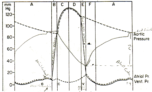
Sorry For The "Scribbling" On This Image...I Wrote On It In Class
Section A is the last 2/3rds of diastole. The a wave on the atrial section is the atrial contraction. This is preceded by the P wave on the ECG. The line of A/B reflects the closure of the AV valves and section B is the isovolumetric contraction phase when the ventricular volume is constant since all the valves are closed on either side of it but the pressure is beginning to rise. This volume constant is the EDV. This is where you hear the S1 heart sound, which is a reflection of the vibrations of the wall of the heart from the sudden stopping of flow and is not the sound made by the closure of the AV valves! Section C – the semilunar valves open when the ventricular pressure exceeds the arterial/aortic pressure. Section C and D are the contraction of the ventricle thus the rapid ejection of blood into the aorta. Section C will be the highest ejection velocity as the generating pressure shoots the blood out once the valves open so initially there is rapid ejection and then in D it will slow down. If the aortic valve was narrow from a stenosis you would hear a murmur loudest when the ejection velocity is greatest so right between S1and S2. The QRS will precede the actual contraction event. The c wave on the atrial trace will be due to ventricular contraction and a back bulge of pressure into the atria. The T wave will occur in section D as the action potentials are repolarizing. Sections B, C, and D all comprise systole. Section E – all four valves are closed. Ventricular volume is constant and at its lowest volume. This is isovolumetric relaxation. This is ESV. The blood has been ejected and no more can go out since the valves are closed. Ventricular pressure is still high so no more can get in either. The atrial v wave will occur here and is the atrial filling phase. The heart sound S2 is heard here and is the vibrations in the wall of the heart after the semilunar valves have closed and the disruption in blood flow. Section F is the rapid filling phase of the ventricles. You could hear an extra heart sound – S3 that would be caused by the vibrations in the walls of the heart during this rapid filling. It sounds very close to S2 and can be synonymous with the sounds in Kentucky. Section A could elicit a 4th heart sound – S4 which is the atrial contraction vibrating if the atria are distended from too much fluid as in early heart failure. This can be synonymous with the sound of Tennessee and comes just before S1. Sections E, F, and A are diastole.
Identify the functional significance of atrial contraction, the origins of the a, c, and v waves of the atrial pressure pulse, and the relationship of the atrial pulse waves to the observable pulsations of superficial neck veins in a supine human subject. Functional significance of the atrial contraction is to squeeze any last bit of blood into the ventricles to give that atrial kick which can constitute an additional 15% of total blood flow. The a,c,v waves are described above. The observable pulsations of the a, c, and v waves can be seen in the jugular vein if the pressures and volumes are elevated here.
Compare and contrast the pressures and flows generated by the right and left ventricles, and describe how these differences in the functional requirements of the two ventricles are reflected in their structure. The RV external work output is normally about one sixth the work output of the LV because of the pressure differences in systolic pressure against which the two ventricles must pump. The RV pressures are normally 15-30/2-8. They must push against the pulmonary pressures which are 15-30/4-12. The LV normal pressures are 100-140/3-12. They must push against systemic arterial pressure which is 100-140/60-90. Consequently the LV is much larger and has a thicker muscular wall than the RV since it must work harder and generate more pressure to push out the blood against a higher pressure gradient.
Compare and contrast the structural and functional characteristics of the atrioventricular and semilunar valves of the heart. The AV valves are the tricuspid (between the right atrium and ventricle) and the mitral (between the left atrium and ventricle.) They have 3 cusps. The AV valves have a thin, filmy nature, which requires almost no backflow to cause closure. The semilunar valves are bicuspid. The pulmonic valve comes between the RV and the pulmonary artery and the aortic valve comes between the LV and the aorta. These valves are much heavier and require rather strong backflow for a few m/sec to close. The all open and close passively when a backward pressure gradient pushes the blood backward, and they open when a forward pressure gradient forces blood in the forward direction.
State the origin of the 1st, 2nd, 3rd, and 4th heart sounds and indicate the timing of these sounds on a diagram of the mechanical events of the cardiac cycle. See objective 6 above.