Physiology I
Section 5
Syllabi
Autonomic Nervous System
Suggested Reading: Guyton Chapters 60
Key Words
Sympathetic (thoraco-lumbar) division: Organized with the peripheral portions of the sympathetic nervous system with two paravertebral sympathetic chains of ganglia that lie to the two sides of the vertebral column, two prevertebral ganglia (the celiac and hypogastric), and nerves extending from the ganglia to the different organs. The sympathetic nerves originate in the spinal cord between the segments T-1 and L-2 and pass from here first into the sympathetic chain and then to the tissues and organs that are stimulated by the sympathetic nerves.
Parasympathetic (cranio-sacral) division:
Parasympathetic fibers leave the central nervous system through cranial nerves
III, VII, IX, and X; the second and third sacral spinal nerves; and occasionally
the first and fourth sacral nerves. About 75% of all parasympathetic nerve
fibers are in the vagus nerves (cranial nerve X), passing to the entire thoracic
and abdominal regions of the body. Therefore, a physiologist speaking of
the parasympathetic nervous system often thinks mainly of the two vagus
nerves. The vagus nerves supply parasympathetic nerves to the heart,
lungs, esophagus, stomach, entire small intestine, proximal half of the colon,
liver, gallbladder, pancreas, and upper portions of the ureters.
Parasympathetic fibers in the third nerve flow to
the pupillary sphincters and ciliary muscles of the eye. Fibers form the
seventh nerve pass to the lacrimal, nasal, and submandibular glands, and fibers
form the ninth nerve pass to the parotid gland.
The sacral parasympathetic fibers congregate in
the pelvic nerves, which leave the sacral plexus on each side of the cord at the
S-2 and S-3 levels and distribute their peripheral fibers to the descending
colon, rectum, bladder, and lower portions of the ureters. Also, this
sacral group of parasympathetics supplies nerve signals to the external
genitalia to cause erection.
Preganglionic fibers: Sympathetic: Comes through the anterior root of the cord into the corresponding spinal nerve. The course of the fibers can be one of the following three: 1): It can synapse with postganglionic neurons in the ganglion that it enters. 2): It can pass upward or downward in the chain and synapse in one of the other ganglia of the chain. Or 3): it can pass for variable distances through the chain and then through one of the sympathetic nerves radiating outward from the chain, finally terminating in one of the prevertebral ganglia.
Parasympathetic: Except in the case of a few cranial parasympathetic nerves, the preganglionic fibers pass uninterrupted all the way to the organ that is to be controlled. Then, in the wall of the organ are located the postganglionic neurons.
Postganglionic fibers: Sympathetic: Originates either in one of the sympathetic chain ganglia or in one of the prevertebral ganglia. From either of these two sources, the postganglionic fibers travel to their destinations in the various organs
Parasympathetic: Short postganglionic fibers, 1 millimeter to several centimeters in length, leave the neurons to spread through the substance of the organ.
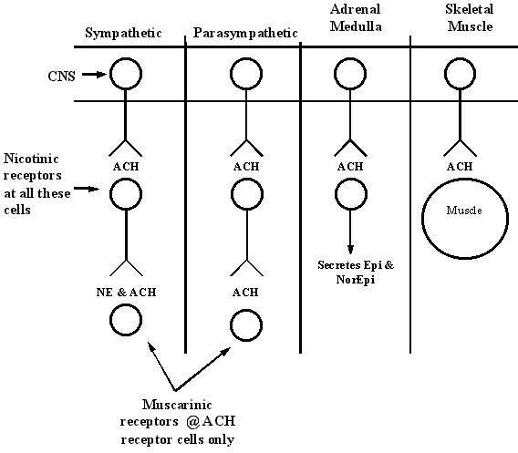
Nicotinic and muscarinic cholinergic receptors: Acetylcholine activates two types of receptors. They are called muscarinic and nicotinic receptors. The muscarinic receptors are found in all effector cells stimulated by the postganglionic neurons of the parasympathetic nervous system as well as in those stimulated by the postganglionic cholinergic neurons of the sympathetic system.
The nicotinic receptors are found in the synapses between the preganglionic and postganglionic neurons of both the sympathetic and parasympathetic systems.

Alpha1 and Alpha2 adrenergic receptors: Excited mainly by Norepinephrine & Epinephrine. See Chart Below.
Beta1 and Beta2 adrenergic receptors: Not excited much by norepinephrine. Excited mainly by Epinephrine. See Chart Below.
| Alpha Receptor | Beta Receptor |
| Vasoconstriction Iris dilation Intestinal relaxation Intestinal sphincter contraction Pilomotor contraction Bladder sphincter contraction |
Vasodilation (B2) Cardio-acceleration (B1) Increased myocardial strength (B1) Intestinal relaxation (B2) Uterus relaxation (B2) Bronchodilation (B2) Calorigenesis (B2) Glycogenolysis (B2) Lipolysis (B1) Bladder wall relaxation (B2) |
Adrenal medulla: Stimulation of the sympathetic nerves to the adrenal medullae causes large quantities of epinephrine and norepinephrine to be released into the circulating blood, and these two hormones in turn are carried in the blood to all tissues of the body. On the average, about 80% of the secretion is epinephrine and 20% is norepinephrine.
Autonomic "tone": The basal rates of activity are known as tone. The value of tone is that it allows a single nervous system to increase or decrease the activity of a stimulated organ.
"Alarm" reaction: Fight or Flight. See Chart below to see what happens
Stress: Mental or physical stress usually excites the sympathetic system, it is frequently said that the purpose of the sympathetic system is to provide extra activation of the body in states of stress )Called the sympathetic stress response). See Chart Below.
|
Effects of Stress or Alarm Reaction
|
Learning Objectives
Describe the general functions and organization of the autonomic nervous system: The autonomic nervous system controls the visceral functions of the body. These include the control of blood pressure, GI motility and secretion, urinary bladder emptying, sweating, body temp, and many other activities. In short it innervates smooth muscle and glands. It is divided into the sympathetic and parasympathetic. There are centers located in the spinal cord, brain stem, and hypothalamus, and portions of the cerebral cortex. The ANS also operates by means of visceral reflexes. That is, sensory signals entering the autonomic ganglia, cord, brain stem, or hypothalamus can elicit appropriate reflex responses directly back to the visceral organs to control their activities. Each sympathetic pathway from the cord to the stimulated tissue is composed of two neurons, a preganglionic neuron and postganglionic neuron. The cell body of each preganglionic neuron lies in the intermediolateral horn of the spinal cord. Once the spinal nerve leaves the spinal canal, the preganglionic neuron can synapse with postganglionic neurons in the ganglion that it enters, pass upward or downward in the chain and synapse in one of the other ganglia of the chain, or it can pass for variable distances through the chain and then through one to the sympathetic nerves radiating outward from the chain, finally terminating in one of the prevertebral ganglia. This differs from the parasympathetic pathway, which has preganglionic fibers that pass uninterrupted all the way to the organ that is to be controlled. Then, in the wall of the organ are located the postganglionic neurons.
Compare and contrast the functional anatomy of the sympathetic and parasympathetic divisions of the autonomic nervous system: As above! The sympathetic pathway has preganglionic synapses near the spinal cord and the postganglionic synapses then travel to the organ being controlled while the parasympathetic pathway has preganglionic synapses that travel all the way to the organ and then synapse with the postganglionic fibers.
Sympathetic (thoraco-lumbar) division: Organized with the peripheral portions of the sympathetic nervous system with two paravertebral sympathetic chains of ganglia that lie to the two sides of the vertebral column, two prevertebral ganglia (the celiac and hypogastric), and nerves extending from the ganglia to the different organs. The sympathetic nerves originate in the spinal cord between the segments T-1 and L-2 and pass from here first into the sympathetic chain and then to the tissues and organs that are stimulated by the sympathetic nerves.
Parasympathetic (cranio-sacral) division:
Parasympathetic fibers leave the central nervous system through cranial nerves
III, VII, IX, and X; the second and third sacral spinal nerves; and occasionally
the first and fourth sacral nerves. About 75% of all parasympathetic nerve
fibers are in the vagus nerves (cranial nerve X), passing to the entire thoracic
and abdominal regions of the body. Therefore, a physiologist speaking of
the parasympathetic nervous system often thinks mainly of the two vagus
nerves. The vagus nerves supply parasympathetic nerves to the heart,
lungs, esophagus, stomach, entire small intestine, proximal half of the colon,
liver, gallbladder, pancreas, and upper portions of the ureters.
Parasympathetic fibers in the third nerve flow to
the pupillary sphincters and ciliary muscles of the eye. Fibers form the
seventh nerve pass to the lacrimal, nasal, and submandibular glands, and fibers
form the ninth nerve pass to the parotid gland.
The sacral parasympathetic fibers congregate in
the pelvic nerves, which leave the sacral plexus on each side of the cord at the
S-2 and S-3 levels and distribute their peripheral fibers to the descending
colon, rectum, bladder, and lower portions of the ureters. Also, this
sacral group of parasympathetics supplies nerve signals to the external
genitalia to cause erection.
Identify the chemical messengers released by pre- and
post-ganglionic fibers of both the sympathetic and parasympathetic divisions of
the autonomic nervous system: See graphic above
In the preganglionic synapse for both sym and para acetylcholine is the
neurotransmitter that is released. These are all nicotinic. At the
postganglionic synapse for the sympathetic either norepi or ach is the
transmitter. These are also nicotinic. At the postganglionic synapse for the
parasympathetic ach is the transmitter and this is muscarinic.
Identify and describe the characteristics and associated second messenger systems of the various types of receptors found on post-synaptic or target organ cell membranes within the autonomic nervous system: The neurotransmitter must bind to a receptor on the outside wall of the cell membrane. This receptor is highly specific and once the transmitter is bound it causes a conformational change to occur in the structure of the protein molecule. The altered protein will either excite or inhibit the cell by either changing the cell membrane permeability or by activating or inactivating an enzyme attached to the other end of the receptor. These enzymes are considered a second messenger. The enzyme is often attached to the receptor protein where the receptor protrudes into the inside of the cell. For instance, binding of epi with its receptor on the outside of many cells increases the activity of the enzyme adenylcyclase on the inside of the cell, and this then causes the formation of cyclic adenosine monophosphate (cyclic AMP). This can in turn initiate any one of many different intracellular actions, the exact effect depending on the chemical machinery of the effector cell.
Identify the general effects of the sympathetic and parasympathetic divisions of the autonomic nervous system and the specific effects of these divisions on the cardiovascular, respiratory, urinary, and gastrointestinal systems:
| Sympathetic | Parasympathetic | |
| Cardiovascular Muscle |
--Increased rate --Increased Force of contraction |
--Slowed rate --Decreased force of contraction (especially of atria) |
| Cardiovascular Coronaries |
--Dilated (B2) --Constricted (Alpha) |
Dilated |
| Respiratory Bronchi |
Dilated | Constricted |
| Respiratory Blood vessels |
Mildly constricted | ? Dilated |
| Urinary Kidney |
Decreased output and renin secretion | None |
| Urinary Detrusor |
Relaxed (slight) | Contracted |
| Urinary Trigone |
Contracted | Relaxed |
| Gastrointestinal Lumen |
Decreased peristalsis and tone | Increased peristalsis and tone |
| Gastrointestinal Sphincter |
Increased tone (most times) | Relaxed (most times) |
Identify the chemical messengers secreted by the adrenal medulla and the functional significance of the adrenal medulla in overall autonomic nervous system actions: Epinephrine and norepinephrine are the chemical messengers from the adrenal medulla. The secretion of epi is the primary one – at 80% while the secretion of norepi is only 20%. The adrenal medulla is an endocrine organ and it does secrete hormones. The cells of the adrenal medulla act as the postganglionic cells. This circulating epi and norepi have almost the same effects on the different organs as those caused by direct sympathetic stim except that the effects last 5-10 times as long because these hormones are removed from the blood slowly. Epi and norepi are usually released from the adrenal medulla at the same time that the different organs are stimulated directly by generalized sympathetic activation. The organs are actually stimulated in two ways and so these two ways can support or substitute for one another.
Describe the general features of medullary, pontine, and mesencephalic control of the autonomic nervous system: These areas control different autonomic functions such as arterial pressure, heart rate, glandular secretion in the GI tract, GI peristalsis, and degree of contraction of the urinary bladder. The most important thing to remember here is that the brain stem controls the arterial pressure, heart rate and respiratory rate. Transection above the brain stem will maintain these three but transection below here will cause these to fall. Another thing to remember is that the autonomic centers in the brain stem act as relay stations for control activities initiated at higher levels in the brain, like the hypothalamus.